Recently, a research project led by University of Michigan- Shanghai Jiao Tong University Joint Institute (UM-SJTU JI) Assistant Professor Jigang Wu has won support from the 2015 National High Technology Research and Development Program of China (863 Program) for young researchers in the area of biomedicine technology. Professor Wu’s project titled “Optical coherence tomography (OCT) and photoacoustic imaging dual-modality endoscopy for diagnosis of atherosclerosis” is a collaboration of his Biophotonics Laboratory with Professor Tian Yang’s Nanophotonics Laboratory, Professor Sung-Liang Chen’s Optical Imaging Laboratory, and Dr. Lixin Jiang’s ultrasound medicine team from Shanghai Sixth People’s Hospital. This is the only 863 youth grant won by SJTU this year, marking a breakthrough in JI’s applied and basic research.
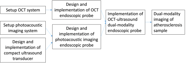
Research flow chart of Professor Wu’s project
It is understood that vascular occlusion of atherosclerotic plaques resulting in rupture and thrombosis accounts for about 50% of human deaths from non-communicable diseases. In recent years, imaging technology, especially in interventional vascular endoscopy, has made considerable progress in early diagnosis, but there is no single existing technique that can fully observe the three main features of plaque breakability, namely thin fibrous cap, lipid core underneath and macrophage activity. As the first effort of JI’s 2013 attempt to build a “multimodal biological imaging and sensing” joint laboratory, this project aims to study OCT- photoacoustic imaging endoscopy, with the design and implementation of its system control and signal processing, in order to fully observe the vulnerable plaques. Through comparative studies with conventional dual-mode ultrasound imaging at the Ultrasound Medicine Department of the Shanghai Sixth People’s Hospital, the team will verify and prepare for transformation of the clinical application.
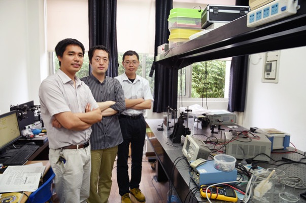
Project team members (from left: Professors. Jigang Wu, Tian Yang, Sung-Liang Chen)
To build a reserve of competitive young researchers (under 35 years of age) with global vision in the biomedical field, the Ministry of Science and Technology set up a biomedical “863 Young Scientists Grant” in 2013, aiming to support biomedical projects in international frontier technology, with the goals to master the international intellectual property rights and core competitiveness, to serve the common needs of life sciences, population health and biomedical industry, and to develop new technologies, new methods, new models and new tools in the biological and pharmaceutical industries. The project “Optical coherence tomography (OCT) and photoacoustic imaging dual-modality endoscopy for diagnosis of atherosclerosis” aims to develop a detecting technique for the rupture of atherosclerotic plaques and thereby provide new prevention and treatment for atherosclerosis. The clinical application is expected to prevent atherosclerotic plaque rupture caused by vascular obstruction and thrombosis, reduce morbidity and mortality related to cardiovascular disease, save significantly in health care costs and improve human health, with significant economic and social benefits.
The 2015 biomedical “863 Young Scientists Project” was very competitive. The committee received 1200 applications from universities, research institutes, hospitals and industries. Through stringent screening, only 50 applicants were finally granted.
Introduction
Dr. Jigang Wu is an assistant professor in electrical engineering at JI. He obtained his B.S. and M.S. in physics from Tsinghua University in 2001 and 2004 respectively. He graduated from California Institute of Technology in 2008 with a Ph.D. in Electrical Engineering. Then he worked as a postdoctoral scholar and research engineer at Caltech for two years. In 2011, he joined JI and has been conducting research in biomedical optical imaging and bio-photonics. He has published 23 papers in Chinese and international journals. He owns seven patents. He has served as reviewers for international journals.
Biophotonics Laboratory(link)
Biophotonics laboratory is dedicated to the development of new biomedical optical imaging methods and explores its application in biological research and medical diagnostics area. Compared with other biomedical imaging technology, optical imaging method is non-invasive and characterized by high resolution and high sensitivity. Current research includes scanning microscopy, focal point of the hole arrays and imaging techniques based on interferometers. The lab is committed to developing new ways to overcome some of the shortcomings of traditional microscopic imaging methods, including the field of view, resolution and sensitivity. The advantages of our technology will make it possible to be applied to health care and other important areas of cancer diagnosis.
Nanophotonics Laboratory(link)
Nanometer-processing technology, high-precision characterization methods and large-scale numerical simulation technology have led to the realization of nano-structures, where the spread and limit of photons are artificially controlled. The magnitude, precision, and efficiency of the interaction between light and matter have been greatly improved. Professor Yang’s team is committed to creating innovative nano-photonics, which will bring about revolutionary change information processing technology, provide efficient and renewable clean energy and make unprecedented biomedical applications possible.
Optical Imaging Laboratory(link)
The lab’s main goal is to develop photoacoustic imaging systems and detect the ultrasonic signal through the optical sensing mode. Compared to the traditional way of using a piezoelectric transducer, the optical sensing helps to improve resolution and signal-noise ratio. In addition, the small dimension of the optical sensor meets the needs of manufacturing a micro-endoscope. It is expected that such a photoacoustic imaging system can improve diagnostic accuracy and the optical ultrasonic detector can open applications in ultrasound-related fields.


