The National Natural Science Foundation of China (NSFC) has announced the 2017 list of funded projects. University of Michigan- Shanghai Jiao Tong University (UM-SJTU JI) Professor Sung-Liang Chen’s “Focusing-free photoacoustic endomicroscopy based on a miniature fiber photoacoustic imaging probe and a mirrored synthetic aperture” is one of the winners.
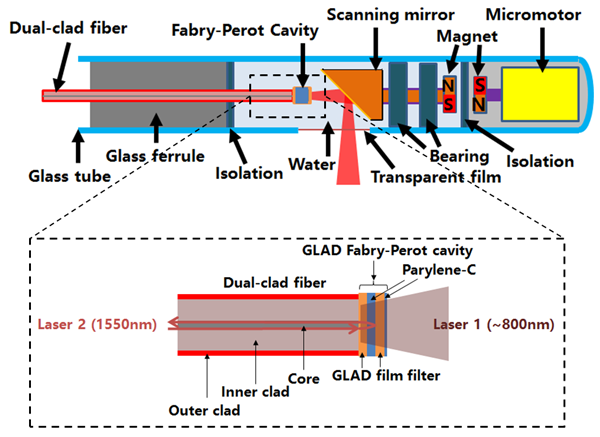 Schematic of the “focusing-free” endoscopic probe
Dashed box: the miniature fiber-optic photoacoustic imaging probe
Photoacoustic imaging is a rapidly developing biomedical technology, where the photoacoustic endoscopy has become a hot research topic in recent years. In acoustic-resolution photoacoustic microscopy, resolutions are typically provided by focused piezoelectric transducers, however, there is a trade-off between resolution and depth of focus (DOF). High-resolution imaging is important for clinical diagnoses, but shallow DOF results in limited observation. It is also unrealistic to adjust the focus of an endoscopic probe to obtain larger DOF in clinical settings. Professor Chen proposed to study “focusing-free” photoacoustic endomicroscopy, using a miniature photoacoustic imaging probe based on a fiber-tip Fabry-Perot optical ultrasound detector, which has advantages, such as small size, broad bandwidth, wide ultrasound receiving angles for reconstructing high-resolution images and a mirrored synthetic aperture, which can be rotated to form a circular aperture that is larger than the size of the endoscopic probe. Together with circular synthetic aperture focusing technique and deconvolution algorithm, high resolution and large DOF can be obtained simultaneously, thus realizing a “focusing-free” photoacoustic endomicroscopy. This project will contribute to applied as well as basic research of photoacoustic endoscopy.
Professor Sung-Liang Chen’s research focuses on three areas:
1. Optical ultrasound sensors: In ultrasound or photoacoustic imaging, piezoelectric transducers are typically used. For some special imaging applications, piezoelectric transducers may have some drawbacks such as bulky size, size-dependent sensitivity, limited bandwidth and limited receiving angles. Chen intends to develop fiber-optic optical ultrasound sensors, which have advantages of miniature size, broad bandwidth and wide receiving angles, etc. Optical ultrasound sensors are advantageous for some specific applications, such as endoscopic imaging. Chen has developed a fiber Fabry-Perot ultrasound sensor with the outer diameter of only 125 µm, the noise-equivalent pressure of 0.4 kPa, the bandwidth of 29 MHz and a −3-dB ultrasound (1−20 MHz) receiving angle of more than ±50°.
Related publications:
[1] G. Li, Z. Guo, and S.-L. Chen, “Miniature all-optical probe for large synthetic aperture photoacoustic-ultrasound imaging,” submitted
[2] D. Cai, G. Li, D. Xia, Z. Li, Z. Guo, and S.-L. Chen, “Synthetic aperture focusing technique for photoacoustic endoscopy,” Opt. Express 25, 20162-20171 (2017)
[3] S.-L. Chen, L. J. Guo, and X. Wang, “All-optical photoacoustic microscopy,” Photoacoustics 3, 143-150 (2015) (Invited Paper)
2. Optical imaging systems: Professors Chen is currently collaborating with Professors Chang-Ching Tu and Jigang Wu. They are working on developing photoacoustic-fluorescence and photoacoustic-optical coherence tomography (OCT) dual-modality and even multimodality optical imaging systems. Photoacoustic imaging has advantages of high contrasts and large imaging depth and is able to acquire rich physiological and pathological information. Fluorescence imaging can utilize fluorescence contrast agents (with an antibody attached) to target an antigen on the cell surface, realizing highly specific targeting. OCT has high resolutions and can be used to image fine structures of tissue. They hope to take advantages of different imaging modalities to obtain more comprehensive diagnostic information.
3. Biomedical photoacoustic imaging: Chen has ongoing collaborations with several hospitals, including Shanghai 6th People’s Hospital (Dept. of Ultrasonography), Renji Hospital (Intensive Care Unit) and Ruijin Hospital (Dept. of Cardiac Surgery). They explore photoacoustic imaging for biomedical and clinical applications. Specific directions include cardiovascular photoacoustic imaging and tumor model photoacoustic imaging, etc.
Related publications:
[1] D. Cai, Z. Li, Y. Li, Z. Guo, and S.-L. Chen, “Photoacoustic microscopy in vivo using synthetic-aperture focusing technique combined with three-dimensional deconvolution,” Opt. Express 25, 1421-1434 (2017)
[2] D. Cai, Z. Li, and S.-L. Chen, “In vivo deconvolution acoustic-resolution photoacoustic microscopy in three dimensions,” Biomed. Opt. Express 7, 369-380 (2016)
Brief Bio of Sung-Liang Chen:
 Sung-Liang Chen is currently an assistant professor at the University of Michigan-Shanghai Jiao Tong University Joint Institute, Shanghai, China. He received his doctoral degree in electrical engineering at the University of Michigan, Ann Arbor, and postdoctoral training at the University of Michigan Medical School. He has published 27 papers in SCI journals such as “Nature Photonics,” “Optics Letters”, “Optics Express,” which have been cited 807 times. His results of the photoacoustic terahertz detection have been reported by mainstream scientific media such as “Gizmag Emerging Technology Magazine,” “IEEE Spectrum,” “Michigan News,” “R & D Magazine,” “Radiology Daily,” and “Science Daily.” His research interests include optical ultrasound sensors, optical imaging systems, and biomedical photoacoustic imaging.
website::http://umji.sjtu.edu.cn/~slchen


